Fibromyalgia: Thoughts About It by a NYC Chiropractor / Applied Kinesiologist/ Neurokinetic Therapist/

Fibromyalgia is a disorder characterized by widespread musculoskeletal pain and sensitivity to touch along with fatigue, problems with sleep and memory and brain fog, People diagnosed with fibromyalgia also often suffer from anxiety, chronic fatigue, depression, irritable bowel syndrome, migraine and tension headaches, painful bladder disorder and TMJ (jaw pain).

According to the Mayo Clinic, “Fibromyalgia can’t be easily confirmed or ruled out through a simple laboratory test. You can’t detect it in your blood or see it on an X-ray. .Because there is no test for fibromyalgia, we must rely solely on your group of symptoms to make a diagnosis.” 

However other blood tests can be taken to rule out conditions like auto -immune, thyroid dysfunction, anemia, viral or bacterial infection, nutritional deficiencies  etc.

One of the first things I look at is the patient’s diet/lifestyle: are they eating processed foods, soda,etc.;drinking enough water;  have a history of medications or antibiotics; are they taking the wrong supplements (too little- too much); do they have  food allergies or intolerance?

It’s not a Food Allergy; maybe it’s a Food Intolerance

I would suspect a leaky gut syndrome and the need to change the person’s diet, test them for allergies/intolerance (igE/IgG)  and ileo-cecal problems and the need for prebiotics and probiotics and other supplements to help repair their gut.

A NYC Applied Kinesiologist on Your Ileo-Cecal Valve (& Houston’s) & Your Health

The Correct Use of Muscle Testing in Nutritional Evaluation in Applied Kinesiology

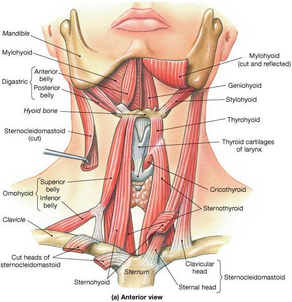
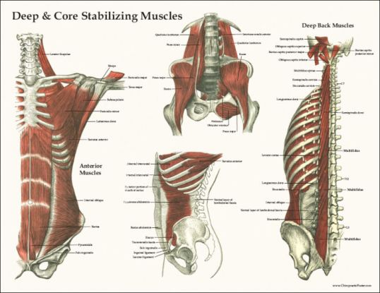
As a Applied Kinesiologist, I test for the function of individual muscles. Are they weak or strong.? The questions to be asked are: why is the muscle weak? Is the muscle on the other side hypertonic or “too stronger.”

As a NKT practitioner, I ask “Is there a dysfunction in the coordination of muscles working in patterns?” I would identify muscle imbalances by using muscle testing to determine what muscles are inhibited and what muscles are compensating (facilitating)for them.

Are these factors causing the muscles to be sensitive to touch?

Is there a problem with the vascular or lymphatic drainage of that muscle?

 I might use neuro-lymphatic , neuro-vascular points and acu-points to increase lymphatic, blood and chi energy to organs and muscles.

How an Applied Kinesiologist Uses Neuro Lymphatics to Improve Health

How a NYC Applied Kinesiologist Uses Neurovascular Pts for Better Physical and Emotional Wellbeing.

Using AK techniques, the flow of energy in the meridians to the organs and muscles can be evaluated and corrected if out of balance. Correction can be made by many methods of stimulation, such as electrical,  small tape patches with metal balls, or simply by mechanically stimulating certain spots.  There is usually an immediate improvement in muscle/organ function after meridian balancing.

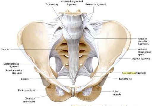
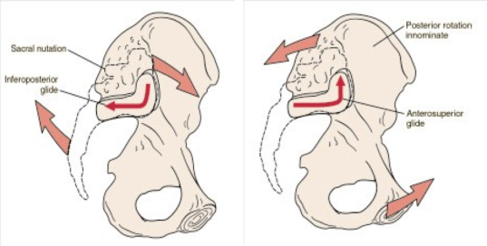
 Is there a problem with the cranial-sacral system? The purpose of the movement of the cranial-sacral system  is to aid the circulation of the cerebrospinal fluid throughout the central nervous system.

Some have suggested that CSF flow along the cranial nerves and spinal nerve roots allow it into the lymphatic channels. Restoring normal cranial-sacral rhythm enables the body to function optimally and may alleviate a wide variety of painful and dysfunctional conditions within the body.

Cranial Sacral Therapy in Applied Kinesiology

Is the patients under chronic stress? Can I help this patient w/ stimulation of their adrenals or with certain supplements?

Is the patient using their diaphragm correctly to order to  get enough oxygen to their body? Stimulation of the diaphragm via spinal adustment and neuro-vasular/neuro-lymphatic/acu-points are done. Patient is taught how to use their diaphragm to get more oxygen into their system.

Can the patient tolerate gentle exercise like chi gong which is is a form of gentle exercise composed of movements that are repeated a number of times, often stretching the body, increasing fluid movement (blood, synovial and lymph) and building awareness of how the body moves through space.?

As a chiropractor, I can use non-force adjustments. It provides a gentle force into the spine of the patient through a very carefully directed and controlled pressure. This method is performed to be able to restore a stronger position and motion to fixated spinal joints taking pressure off the nerves exiting the spinal vertebrae which control the muscles and organs of the body.

These are some of the questions that need to addressed w/ the the complex problem of fibromyalgia; each patient is different and their needs must be addressed.


 © 2018-Dr. Vittoria Repetto

 Want more information on Dr. Vittoria Repetto and her NYC Applied Kinesiology/Chiropractic/ NKT practice at 230 W 13th St., NYC 10011; please go to www.drvittoriarepetto.com

And please check out the Patient Testimonials page on my web site.

 Want to be in the know on holistic information and postings? 

https://www.facebook.com/wvillagechiropracticappliedkinesiologynkt/

Or join me at Twitter: www.twitter.com/DrVRepetto


Hyoid Bone – Its Importance – A NYC Chiropractor/ Applied Kinesiologist / NeuroKinetic Therapist Explains

The hyoid bone  is a horseshoe-shaped bone located in the anterior midline of the neck between the chin and the thyroid cartilage  . At rest, it lies at the level of the base of the mandible or lower jaw  in the front and the third cervical vertebrae(C3) behind.

Unlike other bones, the hyoid is not attached to other bones by muscles or ligaments. The hyoid is anchored by muscles from the anterior, posterior and inferior directions, and aids in tongue movement and swallowing. The hyoid bone provides attachment to the muscles of the floor of the mouth and the tongue above, the larynx below, and the epiglottis and pharynx behind.


 It is located in the front, middle of the throat between the lower jaw and the larynx, which is also called the voice box. The hyoid bone is involved in several functions of the throat, including breathing, swallowing, and talking.

A hyoid bone dislocation occurs when this bone is moved from its normal position in the throat. Common causes of hyoid bone dislocations include car accidents, strangulation, and direct trauma to the neck including surgery for thyroid problems or other anterior neck problems

Symptoms of a dislocated hyoid bone include:  pain, especially when swallowing. difficulty swallowing and talking, swelling of the neck  and severe dislocations may cause difficulty breathing. Intubation for a surgical procedure that is not positioned correctly can affect the position of the hyoid.

The historical importance of the hyoid traces back to the Neanderthals and Homo sapiens for the origin of speech. Unlike in other mammals, the human hyoid bone works together with the tongue and the larynx to produce effective speech as it is responsible for the movement of many small muscles.  So an imbalance in the position of the hyoid and the muscles attachments to the tongue, larynx and pharynx .

One other observation with the hyoid bone is that it can act as one of the body’s gyroscopes; movement of the hyoid bone can affect movement throughout the body and vice versa.  Look at the above picture and imagine the change in position of the hyoid and the cervical (neck) spine if,  for example, there was a break in the rt. clavicle..destroying muscle control of all the muscles on the right. We know of cases from martial arts that some students have problems with their  balance after being kicked in their throats .

Look again at the attachment of muscles above the hyoid and how they attach to the bottom of the jaw  or mandible ; making the hyoid and its muscles part of an examination of TMJ (jaw) problems. 

TMJ Problems (Jaw Problems) and How A Chiropractor/Applied Kinesiologist/NeuroKineticTherapy Practitioner Handles the Problem

As the TMJ is part of the skull and the the cranial bones are checked for alignment ,and we know that the stylohyoid ligament runs from the temporal lobe of the skull to the lesser cornua of the hyoid bone. 

Cranial Sacral Therapy in Applied Kinesiology


Copyright – 2018-Dr. Vittoria Repetto

Addendum: July 21, 2018

New young patient who had flown it from Reno with pain on swallowing…saw my blog. and emailed me….a lot of ant neck muscles, clavicle and cranial adjustments . neck position – Full Ext Pitch and a cervical fixation/bilateral Gluteus Max weakness…I had him getting up down and drinking water in all the different neck positions that were bothering him until he got completely pain-free on swallowing in all range of motions of the neck
I usually enjoy my work but it was great fun !!

Copyright – 2018-Dr. Vittoria Repetto


Want more information on Dr. Vittoria Repetto and her NYC Applied Kinesiology/Chiropractic/ NKT practice at 230 W 13th St., NYC 10011; please go to www.drvittoriarepetto.com

And please check out the Patient Testimonials page on my web site.

 Want to be in the know on holistic information and postings? 

https://www.facebook.com/wvillagechiropracticappliedkinesiologynkt/

Or join me at Twitter: www.twitter.com/DrVRepetto


The Cervical & Vagus Nerve Connection?! – A NYC Chiropractor/Applied Kinesiologist/NeuroKinetic Therapist Explains

I’ve been pondering the possible connection between the Cervical nerve and the Vagus nerve.

The Vagus nerve is the tenth cranial nerve or CN X, and interfaces with parasympathetic control of the heart, lungs, and digestive tract. The Vagus nerve supplies motor parasympathetic fibers to all the organs (except the adrenal glands), from the neck down to the second segment of the transverse colon. The Vagus also controls a few skeletal muscles in the neck or cervical area. The Vagus nerves are paired; however, they are normally referred to in the singular. It is the longest nerve of the autonomic nervous system.

The first and second cervical nerves are responsible for innervation of the head, face, inner middle ear, sinuses, eyes, upper neck, auditory nerves and other areas. The cervical nerves C3-C7 bear the responsibility of innervating the neck, shoulders, thyroid, teeth, tonsils, outer ear, nose, mouth, vocal cords, and more, with some of their individual responsibilities outlined below. The fourth cervical nerve innervates the thoracic diaphragm, leading to the creation of the surgical mnemonic “Cut C4, breathe no more.”

The Vagus Nerve (also known as the “wandering nerve”) exits the upper cervical spine and descends down to the heart and the stomach and the rest of the digestive system. We know of cases where severe massage of the neck muscles affects digestion and respiration.

The Vagus nerve travels along the side of the cervical vertebrae; fibers of the cervical nerves and the vagus innervate similar neck muscles. Misalignment of the upper cervical vertebrae affects the nerves that innervate those muscles. A biofeedback can affect the Vagus.

A relatively new patient made me ponder this possible connection: he had fallen on his head 30 yrs ago. He complained of neck pain, gall bladder pain, low back pain, walking dis-ease, excessive hunger (drinks lots of veggie and fruit juices). He looked emaciated, held his head forward and his shoulders elevated. His X-rays show facet problems w C1-2 vertebra and a slipping forward of C2 on C3; he admits that he self-adjusts his neck.
On examination, found a bilateral weak psoas (a muscle involved in walking) which suggests a fixation of the occipital bone and C1 vertebrae and then also a fixation of C2 and C3.

https://drvittoriarepetto.wordpress.com/2010/12/13/spinal-pain-not-being-helped-see-an-applied-kinesiologist-it-may-be-a-fixation/

I found abnormal sacral movement- a Sacral Wooble, released the erector spinal muscles, adjusted two cranial bones and  worked on acu-points for gall bladder, spleen and liver as well as lymphatic points
His neck’s range of motion is better and walking is not uncomfortable.
Gave instructions on how to track his head back without elevating his chin and told him to stop adjusting his own neck. My dietary advice was to stop juicing and eat solid veggies and more protein.
I emailed him  a youtube video on how to hold his head and another one on the dangers of self-adjustment.

That night and the next 2 days I get emails saying that though he thought I didn’t do a lot; he was not hungry and he had two really good bowel movements and his breathing and his voice is stronger. He was looking forward to his next visit.

Forward lean is a  constant problem with texting, working at a computer and just bad posture. Note that the doctor in the first video mentions that more forward lean, the more years of forward lean, the more medications the patient is on; more stress on cervical…more stress on Vagus?/…more problems w/ organ function.?!!. 

Copyright – 2017-Dr. Vittoria Repetto

Vagal Neurons: New findings in the gut-brain connection

The Spinal Nerve / Autonomic Nerve Connection – A NYC Chiropractor/ Applied Kinesiologist / NeuroKinetic Therapist Explains

Muscles, Connective Tissue Between Organs Influence Illness 


Want more information on Dr. Vittoria Repetto and her NYC Applied Kinesiology/Chiropractic/ NKT practice at 230 W 13th St., NYC 10011; please go to www.drvittoriarepetto.com

And please check out the Patient Testimonials page on my web site.

 Want to be in the know on holistic information and postings? 

https://www.facebook.com/wvillagechiropracticappliedkinesiologynkt/

Or join me at Twitter: www.twitter.com/DrVRepetto

Looking Globally /Not Chasing Pain: A NYC Chiropractor/Applied Kinesiologist/NeuroKinetic Therapist Explains

When patients come in with a local problem such as leg  pain, the first question is where is the source of the pain; is it from the leg itself or the the low back. The other important question is what “set up” the problem. Without answering this question,the problem is not really resolved; the patient will come back with the problem again; you will be just “chasing pain*.”

The answer to the second but more important question may be in the global muscles or core stabilization muscles, these muscles are the Rectus Abdominus, the Transverus Abdominus, the Internal & External Obliques, the Quadratus Lumborum, the Erector Spinae and the Multifidus.

Recently new patient came in complaining of feeling “unbalanced on her right side” siting specifically her right leg and hip.

The lower extremities were a quick fix: a Rectus Femoris – major muscles in the front of the thigh -compensating for a weak Gluteus Maximus. Also the patella and the rotation of the femur (thigh bone) on the tibia (lower leg bone) needed to be adjusted and the Popliteus ( a muscle that rotates the tibia) released.

But during the examination, on lateral flexion of her truck, I noted that her rt shoulder was rotating. I asked her to try to not rotate her shoulder but she was unable; in fact she was unaware of doing it.

I tested her Oblique muscles and they were weak on the right side. Touching (therapy localizing) her Quadratus Lumborum strengthened her Obliques when retested. So I released the QL and the Oblique were strong.

coremuscles


A Category One pelvis which involves rotation was found and fixed.

Low Back Pain & Non Force Adjustments/SOT Blocking: A NYC Chiropractor/Applied Kinesiologist/NeuroKinetic Therapist Explains

When she stood back up, the rt. leg felt balanced and the rotation of her shoulder on lateral flexion was gone.

It was a great illustration of how sometimes part of the problem you have to look above the area of complaint to truly fix the problem; a local problem with a global part.

Stretches and exercises were given as homework.

In my practice, I Iisten, observe and test during the examination and use a combination of NKT, Applied Kinesiology and Chiropractic to help my patients; it’s a strong combination.

How a Combination of Applied Kinesiology, NeuroKinetic Therapy and Chiropractic Works

The Use of Applied Kinesiology in a Chiropractic Examination

© 2017-Dr. Vittoria Repetto

Want more information on Dr. Vittoria Repetto and her  ANYCpplied Kinesiology/Chiropractic/ NKT practice at 230 W 13th St., NYC 10011; please go to www.drvittoriarepetto.com

And please check out the Patient Testimonials page on my web site.

 Want to be in the know on holistic information and postings? 

https://www.facebook.com/wvillagechiropracticappliedkinesiologynkt/

Or join me at Twitter: www.twitter.com/DrVRepetto

*I thank Dr Perry Nickelston for his phrase “Stop Chasing Pain.”

Scars & Muscle Control – A NYC Chiropractor/Applied Kinesiologist/NeuroKinetic Therapist Explains

A while ago I had two patients where scars left from operations affected muscle function.

The first patient came in complaining of acute low back pain that happened on lifting a box that was too heavy for her; one of the muscles I tested is the Rectus Abdominal, it was weak in the clear. The Rectus Abdominal (RA) is part of the support for the Lumbar (low back);she had mentioned that she had had a tummy tuck years ago.
When she touched the scar (therapy localize) which was above her pubes (RA tries into the pubes) her RA tested strong so I massaged the scar and the RA now tested strong.

I find that the T10-T11 vertabrae which are part of the innervation of the RA were subluxated and adjusted them. I also adjusted subluxations in the low back.

The Subluxation/Spinal Joint Dysfunction

I showed her which abdominal exercise I wanted her to do after a daily massage of the scar.

The second patient come in w/ thoracic weakness; she have a history of different cancers.
When I tested her Pectoralis  muscles, both parts (Clavicular & Sternal) tested weak. She said that she was not surprised as a “good part of the muscle was removed” where they removed her breast. I therapy localized the scar from the breast removal and retested as I touched the scar…both parts of the muscle now tested strong.
I massaged the scar… and the muscle tested strong.  I found subluxations at C5 for innervation of the clavicular section and C7 for the sternal section and adjusted them both.
Patient’s homework was to massage that scar before doing exercises for the pectorlis.

Please click on below research articles on relation of scars to muscle pain and function.

CLINICAL IMPORTANCE OF ACTIVE SCARS: ABNORMAL SCARS AS A CAUSE OF MYOFASCIAL PAIN

Surface electromyography of abdominal and back muscles in patients with active scars.

A CASE OF A PATHOGENIC ACTIVE SCAR

Skin, fascias, and scars: symptoms and systemic connections

© 2017-Dr. Vittoria Repetto

This year, I had another patient, an Iraq veteran, who had scars and facial deformity from a gunshot that entered his neck and traveled to the roof of his mouth and the operations to save him.

I used a combination of scar work, cranial sacral and tmj therapy; slowly his face started to change.  His face became more symmetrical and the scars softened. His wife first started seeing the changes after 3 visits and after a few months, his inlaws and fellow veterans saw the difference.

TMJ Problems (Jaw Problems) and How A Chiropractor/Applied Kinesiologist/NeuroKineticTherapy Practitioner Handles the Problem

Cranial Sacral Therapy in Applied Kinesiology


© 2019-Dr. Vittoria Repetto


Want more information on Dr. Vittoria Repetto and her NYC Applied Kinesiology/Chiropractic/ NKT practice at 230 W 13th St., NYC 10011; please go to www.drvittoriarepetto.com

And please check out the Patient Testimonials page on my web site.

 Want to be in the know on holistic information and postings? 

https://www.facebook.com/wvillagechiropracticappliedkinesiologynkt/

Or join me at Twitter: www.twitter.com/DrVRepetto

Medical Articles Of Interest for February 2017

Eggs, High Dietary Cholesterol Not Linked to Increased Risk for Dementia, Alzheimer’s

Dr Repetto’s comment: Egg yolks contain choline, a supplement important for brain health.

Matching the Right Diet to the Right Patient

Sitting Less Linked to Lower Risk of Diabetes

DHA Supplements Linked to Less Progression to Alzheimer’s in APOE4 Carriers

More Support for Gut-Brain Link in Autism

Mediterranean Diet With Olive Oil Boosted HDL Function: PREDIMED

Dietary Protein – From Any Source – May Help Muscle Health

Vitamin D May Protect Against Respiratory Infections

Studies Suggest Cardiovascular Sweet Spot at Two Drinks per Day

Sports Hernias, Adductor Injuries, and Hip Problems Are Linked


Low Back Pain & Non Force Adjustments/SOT Blocking: A NYC Chiropractor/Applied Kinesiologist/NeuroKinetic Therapist Explains

One of the major complaints that causes patients to seek out a doctor of chiropractic is low back pain; it can be caused by either a problem with the lumbar spine or the sacrum.

As a doctor of chiropractic who is also an applied kinesiologist, I use a technique developed by Dr. DeJarnette called Sacro Occipital Technique (SOT); so named because of the relationship between the sacrum (base of the spine) and the occiput (base of the skull).

One of the functions of the sacrum is to pump cerebro-spinal fluid (CSF) from the base of the spine back up the spinal canal to the brain and throughout the nervous system. The occiput also helps to pump CSF. The minute rhythmical motion is essential to optimal health – CSF effectively acts as the circulatory system of the brain and spinal cord.

The pelvis forms the foundational support of the human skeleton. It supports the upper body right up to the skull, and enables us to transfer our weight to our legs. The sacrum is a large bone located at the terminal part of the vertebral spine, where it forms the posterior aspect of the pelvis. The spine holds our body upright, supports all of our organs and provides anchor points for our muscles. It also protects our delicate nervous system. The nervous system controls our body, and can only function normally when our structures are balanced and our pelvis, sacrum and lumbar is stable.

Dr. DeJarnette’s studied two aspects of the sacroiliac joint; the anterior synovial portion and the posterior hyaline cartilage portion. The anterior sacroiliac joint should have motion and this is where sacral nutation and counternutation takes place. The posterior sacroiliac joint is focused on weight-bearing stability and support, which is why at the posterior joint surface there are interlocking of the ridges, and grooves by structures like muscles, ligaments and fascia.

DeJarnette evaluated the weight-bearing characteristics of the sacroiliac joint and determined that when the joint could not adequately support body weight then load bearing stress will be moved upward to the L5/S1 and L4/5 discs, most commonly.

DeJarnette developed an analysis which classified pelvic problems into three different categories and three different non-force techniques using SOT blocks in positions that correct the involved category.

sot-blocks

One of the major complaints that causes patients to seek out a doctor of chiropractic is low back pain; it can be caused by either a problem with the lumbar spine or the sacrum.

As a doctor of chiropractic who is also an applied kinesiologist, I use a technique developed by Dr. DeJarnette called Sacro Occipital Technique (SOT); so named because of the relationship between the sacrum (base of the spine) and the occiput (base of the skull).

One of the functions of the sacrum is to pump Cerebro-Spinal Fluid (CSF) from the base of the spine back up the spinal canal to the brain and throughout the nervous system. The occiput also helps to pump CSF. The minute rhythmical motion is essential to optimal health – CSF effectively acts as the circulatory system of the brain and spinal cord.

Dr. DeJarnette’s studied two aspects of the sacroiliac joint; the anterior synovial portion and the posterior hyaline cartilage portion. The anterior sacroiliac joint should have motion and this is where sacral nutation and counternutation takes place. 

pelvic-ligaments-ant

si-movement

The posterior sacroiliac joint is focused on weight-bearing stability and support, which is why at the posterior joint surface there are interlocking of the ridges, and grooves by structures like muscles, ligaments and fascia.

postsacrummuscles

DeJarnette evaluated the weight-bearing characteristics of the sacroiliac joint and determined that when the joint could not adequately support body weight then load bearing stress will be moved upward to the L5/S1 and L4/5 discs, most commonly.

DeJarnette developed an analysis which classified pelvic problems into three different categories and three different non-force techniques using SOT blocks in positions that correct the involved category.

Category One is a pelvic torsion with altered sacral nutation(motion)  This lack of nutation affects the spinal and cranial meningeal and CSF systems which function to a degree like a closed kinematic chain. Therefore symptoms can be low back pain, chronic shoulder complaints, thoracic outlet syndrome, CSF stagnation, and altered vasomotor function.

Involved muscles can be the piriformis, quadratus lumborum, sacrospinalis, gluteus medius and gluteus maximus. As an applied kinesiologist, I’d check to see if they are hypotonic or hypertonic; as a neurokinetic therapist, I’d check to see if a muscle is weak (or inhibited) by another muscle compensating.

Therapy localization (TL) is done by putting 2 hands on each sacral-iliac joint and then challenging the pelvis for a structural listing and then blocks are put under the patient’s pelvis based to the findings.

The positive Tl’ed side is not adjusted. Cranials are checked.

Category Two happens when ligaments that hold the sacroiliac joint are stretched or sprained, allowing the joint surfaces to separate. Stress can aggravate this ligament weakness via adrenal hormone overdrive.

Symptoms can be low back pain, bowel complaints, possible dysfunction of the reproductive glands and the adrenals, shoulder problems and decreased cervical range of motion.

Involved muscles in addition to the ones mentioned in Category I are the sartorius, gracilis, rectus abdominals and hamstrings along with the iliolumbar ligament. These structures are tested via applied kinesiology and neurokinetic protocols mentioned above. Cranials again are checked.

Category Three occurs when the low back can no longer tolerate the physical stressors placed on it and involves both disc and nerve root aggravation. This can be a sudden one-off event such as a lift, or it can be a pre-existing weakness that is aggravated. Often Category Three produces pain in the low back and sometimes pain radiates down a leg as sciatica.

Muscles to be checked are the psoas as it attaches into the front of the lumbar vertebrae as well as the muscles involved in Category One as Category Three can be a Category One that was never corrected.

Correction in all the categories involves using blocks under the pelvis in specific directions related to the category and the subluxation/misalignment of the pelvis/sacrum. The patient’s weight and breathing help to balance the low back, sacrum and CSF flow and takes the pressure or irritation off the nerve. This allows the body to heal.

Patient is told to ice the involved areas, how to do daily activities, given stretches and exercises and advised on nutrition to help the body heal.

For more information on issues mentioned:

Cranial Sacral Therapy in Applied Kinesiology

How a Combination of Applied Kinesiology, NeuroKinetic Therapy and Chiropractic Works

The Use of Applied Kinesiology in a Chiropractic Examination

© 2017-Dr. Vittoria Repetto

Want more information on Dr. Vittoria Repetto and her NYC Applied Kinesiology/Chiropractic/ NKT practice at 230 W 13th St., NYC 10011; please go to www.drvittoriarepetto.com

And please check out the Patient Testimonials page on my web site.

 Want to be in the know on holistic information and postings? 

https://www.facebook.com/wvillagechiropracticappliedkinesiologynkt/

Or join me at Twitter: www.twitter.com/DrVRepetto

Medical Articles of Interest for December 2016

Gut Microbiome Again Linked to Parkinson’s Symptoms

High Dietary Magnesium Intake Tied to Less Stroke, Diabetes, Heart Failure

Muscle Strength Gains Linked to Better Brain Function

Gastric Acid Blockers Boost Risk of Iron Deficiency

The Gut–Brain Connection

Statin Use Linked to Increased Parkinson’s Risk

Chronic Use of Proton Pump Inhibitors Increases Heart Risk

Selenium Supplement Role Unclear in Autoimmune Thyroiditis

Sauna Use Linked to Lower Dementia, Alzheimer’s Risk


Want more information on Dr. Vittoria Repetto and her NYC Applied Kinesiology/Chiropractic/ NKT practice at 230 W 13th St., NYC 10011; please go to www.drvittoriarepetto.com

And please check out the Patient Testimonials page on my web site.

 Want to be in the know on holistic information and postings? 

https://www.facebook.com/wvillagechiropracticappliedkinesiologynkt/

Or join me at Twitter: www.twitter.com/DrVRepetto


Anterolateral Lower Leg Pain &/or Foot Drop – Peroneal Nerve Entrapment – A NYC Chiropractor /Applied Kinesiologist /NeuroKinetic Therapist Explains

Occasionally a patient comes in with pain along the anterior lateral part of their lower leg and they may also have difficulty lifting the top of their foot upward at the ankle (dorsiflexion) or moving the foot outward at the ankle (eversion). This also affects the patient’s gait (ability to walk properly)

movements-of-foot

They may have been told that their problem is sciatica since branches of the sciatic nerve goes all way down the leg and into the toes. They may have been to a physical therapist or another chiropractor who worked on the low back where the sciatic nerve originates without any relief.

How a NYC Chiropractor /Applied Kinesiologist /NeuroKinetic Therapist Treats Sciatica

But the problem is the peroneal nerve which branches off the sciatic just below the knee; it goes from the common peroneal nerve which starts at the lateral back of the knee near the inner side of the biceps femoris  (part of the hamstings), goes to the back of the fibula bone and then branches off to the superficial peroneal nerve and the deep peroneal nerve.

The deep peroneal nerve innervates the muscles of the anterior compartment of the leg which are: tibialis anterior, extensor hallucis longus, extensor digitorum longus, and the peroneus tertius. Together these muscles are responsible for dorsiflexion of the foot and extension of the toes.

The superficial peroneal nerve passes between the peroneus muscles and the extensor digitorum longus, and goes into the deep fascia at the lower third of the leg; it supplies the muscles of the lateral compartment of the lower leg : peroneus longus and peroneus brevis. These assist with eversion and plantar flexion of the foot.

The deep peroneal nerve passes inferior and medially, deep to extensor digitorum longus,  and comes into relation with the anterior tibial artery above the middle of the leg; it then descends with the artery to the front of the ankle-joint, where it divides into a lateral and a medial branch. The deep peroneal nerve innervates the muscles of the anterior compartment of the leg which are: tibialis anterior, extensor hallucis longus, extensor digitorum longus, and the peroneus tertius. These muscles are responsible for dorsiflexion of the foot and extension of the toes.

superficial-and-deep-peroneal-nerve-and-muscles

Spasms of muscles along the route of these nerves can cause nerve entrapment and result in loss of muscle function or pain along the route of the nerves. As muscle attaches to bones and joints, spasms can cause misalignments of these structures and this can further complicate the problem.

Sometimes the muscle spasms are complicating for a muscle that is inhibited somewhere along the lateral or posterior kinetic chain such as the gluteus maximus, gluteus medius, the hamstrings and the rectus femoris or the psoas which overwork in order to to lift the leg to clear the dropped foot during walking. Neurokinetic therapy works well answering this question and correcting the problem.

As a applied kinesiologist and neurokinetic therapist I will either place my finger or hand on specific areas and then retests the muscle to see if the weakness is corrected. This is called therapy localization.

If the therapy localization is positive and the area involved is a joint or a muscle, I can move the area in a way to stimulate neuro/mechano receptors in the joint or muscle. This is called a “challenge’ and shows the direction of manipulation needed to improve function of the joint or muscle.

As a chiropractor, I would not only adjust the involved joints but check to see if the change in gait muscles has affected the spine and adjust affected level.

Stretches are given to the previously complicating muscles and exercises given to the previously inhibited muscles in order to break the pattern that caused the problem.

For  more information, please read:

The Use of Applied Kinesiology in a Chiropractic Examination

How a Combination of Applied Kinesiology, NeuroKinetic Therapy and Chiropractic Works


© 2016-Dr. Vittoria Repetto

Want more information on Dr. Vittoria Repetto and her NYC Applied Kinesiology/Chiropractic/ NKT practice at 230 W 13th St., NYC 10011; please go to www.drvittoriarepetto.com

And please check out the Patient Testimonials page on my web site.

 Want to be in the know on holistic information and postings? 

https://www.facebook.com/wvillagechiropracticappliedkinesiologynkt/

Or join me at Twitter: www.twitter.com/DrVRepetto


Medical Articles of Interest for October 2016

SSRIs Disrupt Sleep in the Elderly, May Contribute to Dementia

Phytoestrogens May Benefit Health but Also Prompt Concern

Unclear If Sports Raise Later Arthritis Risk 

Sleep Duration Important in Weight Management

Micronutrient Deficiency Often Unnoticed in PPI Users

” PPIs have been associated with Clostridium difficile infection and with micronutrient deficiency. “Our hypothesis was that even though gastroenterologists know about micronutrient deficiency, they aren’t looking for it,” he explained. The researchers reviewed the electronic health records of 41 patients with Barrett’s esophagus who were on long-term PPI therapy to see if their levels of vitamin B₁₂, ferritin, or magnesium were ever tested”

Aerobic Exercise May Provide ‘Small’ Improvement in Vascular Cognitive Impairment

IBS: Gut Bacteria May Predict Who Benefits From FODMAP Diet

“Patients who did not respond to the low-FODMAP diet had more severe dysbiosis at baseline than responders. The traditional diet had no effect on overall bacterial composition, but in the FODMAP group, there was a significant reduction in potentially beneficial Bifidobacterium. This was more prominent in nonresponders than in responders.”

New Guideline Says Calcium Safe From CVD Standpoint

(Dr. Repetto’s comment – One should take calcium with magnesium, vitamin D and vitamin K. One should aim to get 1200 mg/day and half of that from food.)